While most people are familiar with the visual signs of skin cancer, the microscopic world reveals a fascinating and crucial aspect of diagnosing this disease.
Understanding skin cancer at the microscopic level empowers dermatologists like those at the Center for Dermatology and Laser Surgery to make informed decisions about your care. Keep reading to learn more about skin cancer and what our dermatopathologists are looking for when they receive a skin sample from a biopsy.
What Is a Biopsy for Skin Cancer?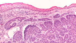
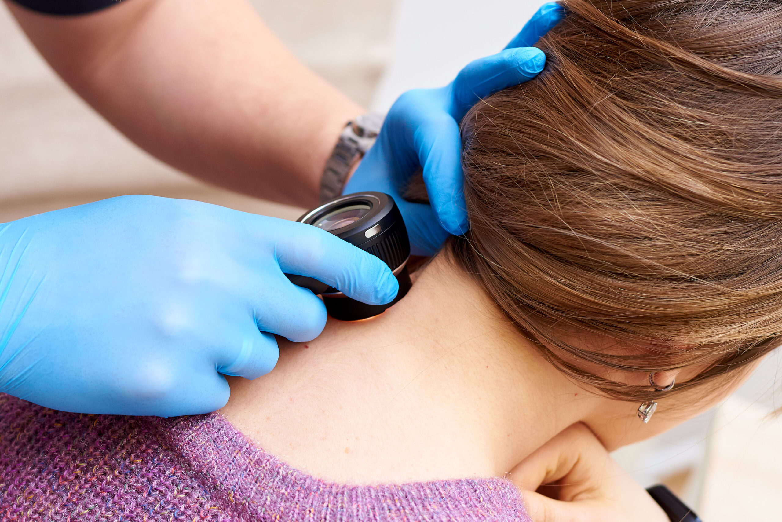
The journey to a skin cancer diagnosis often begins with a visual examination by a qualified dermatology provider. During this examination, your provider will assess the mole or lesion for key characteristics associated with skin cancer, such as asymmetry, border irregularity, color variation, diameter, and evolution (ABCD rule).
Dr. Emily Green, Director of Golden State Dermatology’s Pathology Lab explains how microscopic analysis plays a vital role in accurate skin cancer detection and treatment: “When a dermatopathologist examines biopsy slides, she can observe the individual cells and their organization. In cases of cancer, the cells may appear disorganized. The features visible under the microscope enable the personalized ordering of additional tests, leading to the most accurate diagnosis.”
Sometimes, a visual examination alone cannot definitively confirm or rule out skin cancer. If your provider suspects a potential malignancy, the next step typically involves a biopsy.
A biopsy is a minimally invasive procedure where a small tissue sample is extracted from the suspicious area. This sample is then sent to a pathologist at our Dermatopathology Lab for microscopic examination.
What Should I Look For?
While a dermatopathologist will be looking for specific signs under a microscope, you may also see signs that are viewable. Early detection is key to successful skin cancer treatment. It’s important to be aware of the warning signs and perform regular skin self-exams. Here are some characteristics to look for:
Asymmetry: One half of a mole or lesion does not match the other half.
Border irregularity: The edges of a mole or lesion are irregular, ragged, notched, or blurred.
Color variation: The color of a mole or lesion varies from one area to another, with shades of brown, black, tan, white, red, or blue.
Diameter: The diameter of a mole or lesion is larger than 6 millimeters (about the size of a pencil eraser).
Evolving: A mole or lesion that changes in size, shape, color, or texture over time.
If you notice any of these changes in a mole or lesion, it’s essential to consult with a dermatologist for a professional evaluation. Early detection and prompt treatment can significantly improve the outcome of skin cancer.
How Are Cancer Cells Different Under a Microscope?
A pathologist utilizes a powerful microscope in addition to specific preparations to the specimen to examine the cells within the biopsied tissue. Healthy skin cells exhibit a well-defined, organized structure.
In contrast, cancerous cells often display abnormalities that can be detected under the microscope. Here’s a breakdown of some key microscopic features associated with different types of skin cancer:
Basal Cell Carcinoma (BCC)
The most common form of skin cancer, BCC, typically appears under the microscope as clusters of tightly packed, round, or oval-shaped cells with darkly stained nuclei.
Squamous Cell Carcinoma (SCC)
This skin cancer originates in the squamous cells of the outer layer of the epidermis. Under the microscope, SCC can appear as sheets of atypical cells with abnormal shapes and sizes.
These cells may also exhibit increased mitotic activity, indicating a higher rate of cell division.
Melanoma
The most aggressive form of skin cancer, melanoma, presents unique microscopic features. Pathologists look for cells with irregular shapes and sizes, prominent nucleoli (the dark center of the cell nucleus), and signs of invasion into deeper layers of the skin.
What Are Subtypes and Grading?
Microscopic examination allows not only the identification of skin cancer but also further classification into subtypes. Each subtype possesses specific characteristics that can influence treatment decisions.
For example, certain types of BCC may require more aggressive surgery than others. This helps determine the grade of the cancer.
The grade refers to the aggressiveness of the cancer cells and their growth rate. Lower-grade cancers typically grow slower and are less likely to spread compared to higher-grade cancers.
 Why Is Early Detection Important?
The microscopic analysis of skin cancer plays a critical role in early detection, which is crucial for achieving optimal treatment outcomes. When skin cancer is identified and addressed in its early stages, the chances of successful treatment are significantly higher.
Regular skin cancer screenings by your doctor at the Center for Dermatology and Laser Surgery are vital for early detection.
If you have any concerns about your skin or have noticed any suspicious moles or lesions, schedule an appointment with your doctor at the Center for Dermatology and Laser Surgerytoday. Early detection and prompt treatment are crucial for optimal outcomes.
Schedule a Consultation Today
Do you have any concerns about your skin health? Schedule a skin cancer screening at the Center for Dermatology and Laser Surgery today.
Our team of experienced dermatology providers is dedicated to providing comprehensive skin care solutions. By prioritizing skin health, you can take proactive steps to protect yourself from skin cancer and maintain a healthy, radiant complexion.